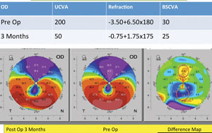

7, 9 These cases have been defined in some series as “pellucid-like keratoconus” (PLK), 7 being considered as a different presentation of keratoconus. Those cases presenting a crab claw-like pattern in the anterior corneal topographic map, but without associated peripheral thinning, cannot be diagnosed as true PMD. 7, 8 Additional tools must be used for an adequate diagnosis of PMD, such as slit-lamp examination and pachymetric data, allowing the clinician to identify the position of the point of MCT. 6 However, this specific topographic pattern may also be present in keratoconus, easily being misdiagnosed as a true PMD in clinical practice. 4Ĭlassic manifestation of PMD is flattening of the vertical meridian above the thinning band, inducing an area of steepening below this band, an “against-the-rule” astigmatism, 5 and a crab claw-like topographic pattern. 3 Furthermore, PMD seems to progress slower than keratoconus, presenting less visual alteration over time. 1, 2 It is considered as a rare condition 1 and is usually diagnosed in the later decades of life compared to keratoconus. Pellucid marginal degeneration (PMD) is a noninflammatory and progressive ectatic corneal disease characterized by a narrow band of corneal thinning 1–2 mm from the limbus (commonly inferior limbus). Keywords: Corneal ectasia, Keratoconus, Keratoconus progression, Pellucid marginal degeneration.

The presence of two different conditions in the same patient (case 1) and the progression from inferosuperior asymmetry to the development of a crab claw-pattern (case 2) suggest that PMD, PLK, and keratoconus may be different phenotypic presentations of the same pathophysiological condition. The second case presented was a 19-year-old male without clinical signs of ectasia at baseline that progressed to PLK with evident changes in topographic and pachymetric maps but maintaining the point of minimum corneal thickness (MCT) in the central area. The first case was a 58-year-old male who developed a true pellucid marginal degeneration (PMD) in one eye and with a nonprogressive PLK in the other eye. Currently, according to clinical examination, these two cases are diagnosed as pellucid-like keratoconus (PLK). We report the long-term follow-up of two cases of untreated corneal ectasia presenting a crab claw-like sagittal and tangential topographic pattern at baseline, but without peripheral thinning. Source of support: The author David P Piñero has been supported by the Ministry of Economy, Industry and Competitiveness of Spain within the program Ramón y Cajal, RYC-2016-20471. Differences in the Long-term Progression Course of Two Cases of Pellucid-like Keratoconus: Are they the Same Condition with Different Phenotypic Presentation? Int J Kerat Ect Cor Dis 2019 8(1):29–33. International Journal of Keratoconus and Ectatic Corneal Diseases Volume 8 | Issue 1 | Year 2019ĭifferences in the Long-term Progression Course of Two Cases of Pellucid-like Keratoconus: Are they the Same Condition with Different Phenotypic Presentation?Īntonio Martínez-Abad 1, David P Piñero 2ġDepartment of Research and Development, Vissum Corporation, Alicante, Spain 2Department of Optics, Pharmacology and Anatomy, Group of Optics and Visual Perception, University of Alicante, Alicante, SpainĬorresponding Author: David P Piñero, Department of Optics, Pharmacology and Anatomy, Group of Optics and Visual Perception, University of Alicante, Alicante, Spain, Phone: +34-965903500, e-mail: to cite this article Martínez-Abad A, Piñero DP.


 0 kommentar(er)
